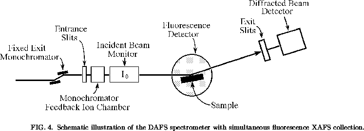
The DAFS experiments described in this chapter were performed at the National Synchrotron Light Source (NSLS), using beamline X23A2. The synchrotron radiation source provided an intense continuous energy spectrum, strong collimation, small source size, and a high degree of polarization. An XAFS beamline was chosen because rapid and precise energy scanning is essential for DAFS measurements. The beamline was adapted by adding a 2-circle spectrometer to give it diffraction capabilities. The 2-circle spectrometer had limited reciprocal space coverage, but it was sufficient for all the experiments described in this chapter. All of the work reported here was done using specular Bragg reflections. The configuration of the X23A2 beamline during the DAFS experiments is shown in Fig. 4.
In the fixed exit monochromator, Si(220) crystals were used for the Cu
measurements and Si(311) crystals were used for the
In0.2Ga0.8As
and
YBa2Cu3O6.6
measurements. The
energy calibration procedure is described in reference
[21]. During the DAFS experiments, the beamline produced
a measured flux of about ![]() photons per second in a 1 mm high
by 5 mm wide beam, with a measured energy spread of about 2 eV.
photons per second in a 1 mm high
by 5 mm wide beam, with a measured energy spread of about 2 eV.
The Bragg diffracted radiation was selected with a 3 mm high by 5 mm wide slit located 27 cm from the sample. This provided adequate suppression of the fluorescence from the sample while accepting essentially all of the diffracted beam. At each energy, the diffractometer was adjusted to keep the momentum transfer fixed; to obtain reliable intensity measurements we found it essential to accurately track the Bragg peak versus energy. For the thin epitaxial film samples we studied, which had broad and smooth mosaic distributions, the peak intensities were proportional to the integrated intensities, and we report here our peak intensity measurements.
We used integral (current mode) techniques to maximize the number of
detected photons. Because commercial NaI(Tl) scintillation detectors can
only count up to about ![]() photons per second and the samples in
this study often had diffracted intensities greater than
photons per second and the samples in
this study often had diffracted intensities greater than ![]() photons per
second, both the incident and diffracted x-ray intensities were measured
using ionization chambers. The noise, set by photon counting statistics,
is proportional to the square-root of the number of detected photons and
should be less than the fine structure signal. Since the fine structure
signal can be as low as
photons per
second, both the incident and diffracted x-ray intensities were measured
using ionization chambers. The noise, set by photon counting statistics,
is proportional to the square-root of the number of detected photons and
should be less than the fine structure signal. Since the fine structure
signal can be as low as ![]() , at least
, at least ![]() detected photons are
required just to reduce the noise to the level of weak fine structure
signals. To reduce the sensitivity of the signal and monitor ionization
chambers to the second or third harmonics of the 9 Kev fundamental, the gas
in each ion chamber was chosen to produce an absorption of about 50% at 9
Kev.
detected photons are
required just to reduce the noise to the level of weak fine structure
signals. To reduce the sensitivity of the signal and monitor ionization
chambers to the second or third harmonics of the 9 Kev fundamental, the gas
in each ion chamber was chosen to produce an absorption of about 50% at 9
Kev.
To compensate for the incident intensity variations, we divided the diffracted intensity at each energy by the incident beam monitor signal. The fluorescence from the sample was measured simultaneously with the DAFS signal using a 10 cm diameter ionization chamber [22]. This measured fluorescence signal was used as an energy reference and to allow comparisons of the XAFS and DAFS signals. The measured fluorescence was also used to calculate the absorption corrections for the Cu metal and YBa2Cu3O6.6 samples.