Overview l Microprobe Characteristics l Microprobe Experiments l Microtomography Characteristics l Microtomography Experimments l Design Team
![]()
Overview
l Microprobe Characteristics l Microprobe Experiments l Microtomography
Characteristics l Microtomography Experimments
l Design Team
The x-ray microprobe instrumentation is designed to be capable of fluorescence analysis (XRF), XAFS and computed microtomography (CMT). The undulator microprobe will be designed for ultrahigh spatial resolution, highest sensitivity and pencil-beam microtomography. The microprobe experiments will require < 10
mm synchrotron beams which will be produced with microfocusing Kirkpatrick-Baez mirrors. This capability is crucial for determination of elemental partitioning and speciation determinations at natural concentrations. The Kirkpatrick-Baez optics have the advantage of large working distances (50 mm from the downstream end of the second mirror), achromatic operation, i.e. no refocusing is required as the monochromator energy is scanned, and fixed offset. Microprobe Characteristicsl
Goal is 1 micron beam with ppb sensitivity for trace elementsl
Compositions of buried components (fluid inclusions)l
Compositional mapping (diffusion, zonation)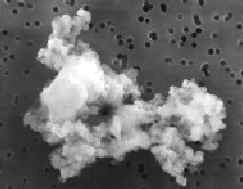 |
Electron image of an interplanetary dust particle (~10 microns) collected in the Earth's stratosphere. These particles derive from asteroids and comets and are aggregates of silicates, carbonaceous material,oxides and hydrous phases such as smectite. Detailed compositional analyses provides direct information on the nature of these primitive solar system bodies. Courtesy Jet Propulsion Laboratory. Copyright (c) California Institute of Technology, Pasadena, CA. |
Microtomography Characteristics
l CAT scans with micrometer resolution
l Elemental
specificity using fluorescence and edge tomography
l High
energy needed for large samples
l Environmental
chambers
l Near real
time visualization
l Dynamic
studies of fluids in rocks and soils (e.g. diffusivity)
l Root-soil-micro-organism
interactions
l Microstructure
visualization of rare and precious objects (meteorites, fossils, ice cores)
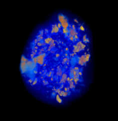 |
The micro- tomography image is of a meteoritic (chondrite) pebble (5 mm in diameter) collected from MacAlpine Hills, Antarctica. Tomographic imaging is of interest for determining locations and size distributions of clasts, fusion crust thickness, modal abundance of phases,porosity, density, and possibly the distributions of terrestrial weathering products. The ability to determine the locations of clasts non-destructively is valuable, for example, in developing sampling strategies and allocation plans for rare meteorites such as those from the Moon and Mars. |
| Steve Sutton, Chairman University of Chicago, Building 434A, Argonne National Laboratory, 9700 South Cass, Argonne, IL 60439; phone : 630-252-0426; fax : 630-252-0443, sutton@cars.uchicago.edu |
|
| Mark Rivers University of Chicago, Building 434A, Argonne National Laboratory, 9700 South Cass, Argonne, IL 60439; phone : 630-252-0422; fax : (630) 252-0443, rivers@cars.uchicago.edu |
|
|
Darrell Schulze Agronomy Department, Purdue University, 1150 Lilly Hall of Life Sciences, W. Lafayette, IN, 47907; phone : 317- 494-8062, fax : 317-494-6508, dschulze@dept.agry.purdue.edu |
|
Keith Jones Department of Applied Science, Building 815, Brookhaven National Laboratory, Upton , NY, 11973; phone : 516- 282-4588, fax: 516-282-7905, jones@bnlx26.nsls.bnl.gov
|
| Ian Steele University of Chicago, Department of Geophysical Science; phone : 773-702-8109, fax : 773-702-9505, steele@geo1.uchicago.edu
|
Overview l Microprobe Characteristics l Microprobe Experiments l Microtomography Characteristics l Microtomography Experimments l Design Team