High-spin to low-spin transitions in FeO and Fe2O3 probed by high-resolution x-ray fluorescence spectroscopy
Badro, J, Hemley, R J, Mao, H, Struzhkin, V V, Shu, J, Shen, G, Kao, C, Rueff, J, Hu, J
As an end-member mineral of the Earth's deep mantle and as a possible component of the Earth's core, the bonding and the electronic structure of wustite (FeO) are of considerable geophysical importance. To this purpose, we used high resolution x-ray fluorescence technique at the Fe K-beta edge to probe the spin state of the system, and this for the first time in the diamond anvil cell. Because of the relatively high absorption of electromagnetic radiation by diamond at these energies, the x-ray beam was directed through a high strength beryllium gasket. The experiments were conducted at the Advanced Photon Source at Argonne National Laboratory. We used a collimated 10x4 microns (vertical x horizontal) white beam on the undulator insertion device beamline at sector 13, Consortium for Advanced Radiation Sources of the University of Chicago. The transition is probed by the disappearance of the low energy satellite structure in the emission spectrum resulting from the 3p core hole---3d exchange interaction in the emission final state. At room temperature, FeO remains in rhombohedral phase up to at least 150 GPa. A high-spin to low-spin transition in FeO has been reported by Mossbauer spectroscopy for pressures between 80 and 120 GPa. The high resolution x-ray fluorescence technique indicate that the system persists in the high-spin state to at least 150 Gpa. A low-spin state was detected, however, after the sample was laser heated at 150 GPa. A transition from a high-spin to low-spin state is observed to occur in Fe2O3. This compound undergoes a structural phase transition (hematite to perovskite phase) at 50 GPa. A measurement at 72 GPa indicates that the system is in low-spin state.
Experiment
Four important components of the XES experiment are:
Important high-pressure cell modification (the use of Be gasket) allowed the first measurement of low-energy X-ray signal.
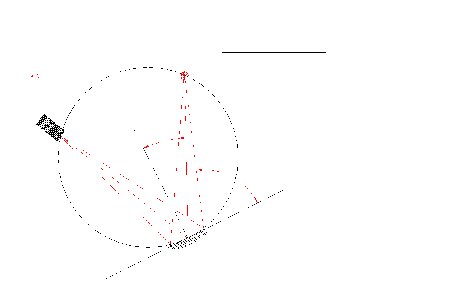
Fig.1. Experimental setup for high resolution XES. Analyzers used were Ge and Si crystals.
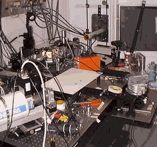
Fig. 2. The setup at the APS, Chicago
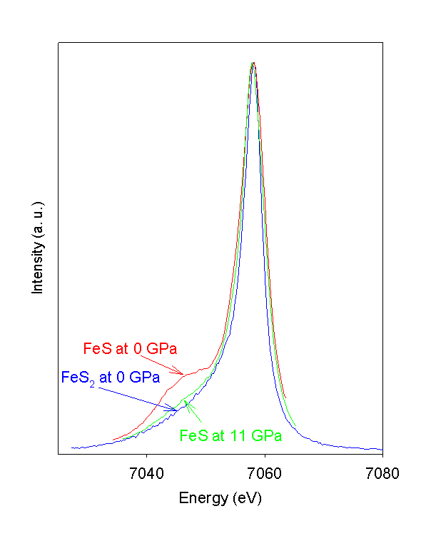
Fig. 3. (a) K
b emission spectra of FeS during compression to 11.5 GPa ,(b) FeS sample, decompression,
(c) comparison with FeS2 (low-spin configuration) emission. 11 GPa emission spectrum of FeS is analogous to low-spin FeS2 emission.
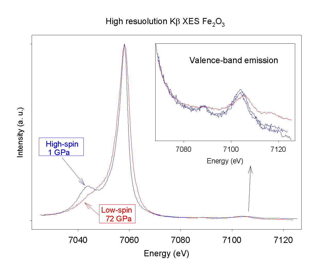
Fig. 4. K
b emission spectra of Fe2O3 at 1 at 72 GPa, clearly showing low spin state at 72 GPa. The change to low-spin state is evident, however, small shoulder is visible in the satellite region closer to main peak in the low-spin state spectrum.
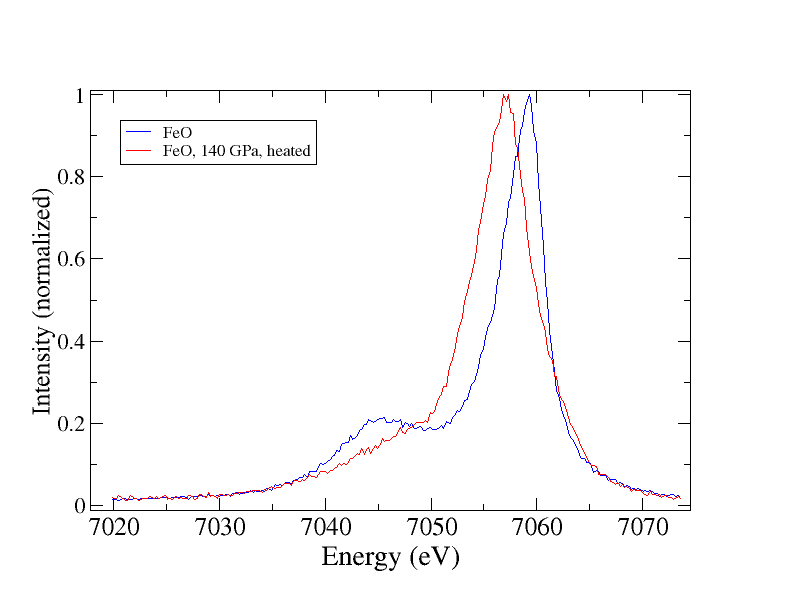
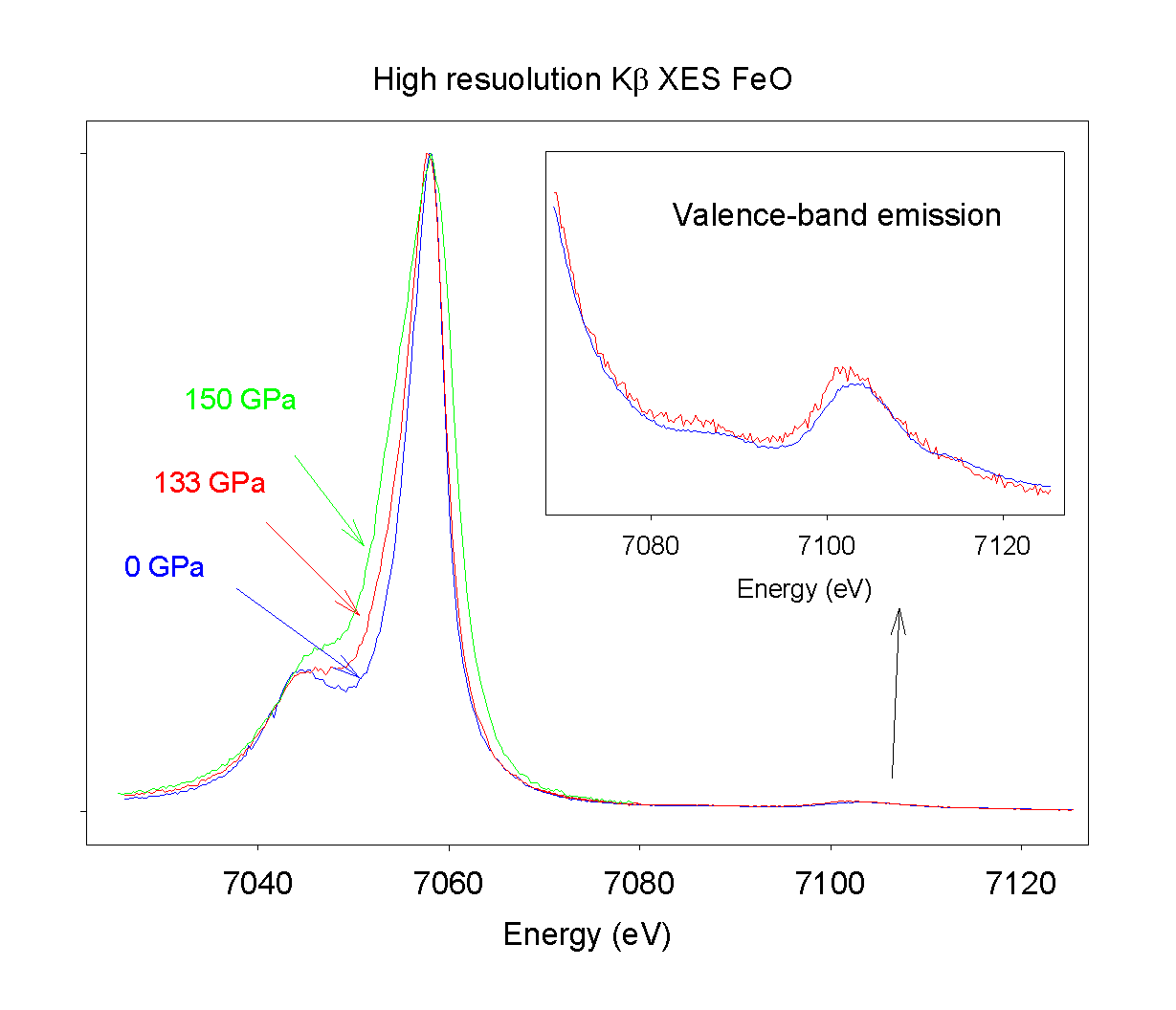
Fig. 5. K
b emission spectra of FeO up to 150 GPa. The spectra show high-spin state all the way up to 150 GPa at room temperature. With the laser heated sample at 140 GPa, a low-spin state was detected. These results do not agree with Mössbauer data [1]. At 150 GPa there is probably a contribution from the sample part residing at lower pressures, due to defocused beam (damaged focusing KB mirrors) and pressure gradients in the sample. However, similar pressure gradient problems are also present in Mössbauer experiments [1].